Bachelor of Diagnostic Imaging (Radiography)
Overview
Job Growth
N/A
Duration
N/A
Avg. Salary
N/A
Career Paths
N/A
Program Description
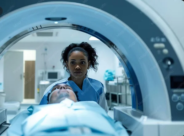
The Bachelor of Diagnostic Imaging (Radiography) trains students to produce and interpret medical images that help diagnose disease and guide treatment. You will study anatomy, physiology, medical physics, radiographic techniques, ultrasound, computed tomography, image processing, radiation safety, and patient care. The program combines classroom theory with hands-on training in imaging equipment and clinical placements in hospitals. Graduates can work as radiographers in public and private hospitals, diagnostic centres, clinics, or continue to specialize in modalities like CT, MRI, or sonography. This program builds practical clinical skills and opens doors to stable, respected careers in healthcare and medical science.
Aims & Objectives
Develop competent clinical skills in acquiring diagnostic images using X-ray, ultrasound, and CT equipment, measured by supervised clinical assessments.
Master radiation protection principles and safe operation of imaging equipment, demonstrated through practical exams and radiation safety logs.
Understand human anatomy and pathology relevant to imaging, assessed by written and practical anatomy tests.
Produce accurate medical documentation and radiographic reports, evaluated through case-based assignments and portfolio reviews.
Why Choose This Program?
High demand for diagnostic services
Hospitals and clinics in Ghana and beyond need trained radiographers, offering steady employment in public and private sectors.
Hands-on, practical learning
Strong emphasis on clinical placements and equipment training gives real-world experience before graduation.
Clear career pathways
Opportunities to specialize in CT, MRI, ultrasound, or pursue roles in management, research, or education.
Impactful patient care role
You play a key role in diagnosis and treatment, working closely with doctors and other health professionals to improve patient outcomes.
Skills & Tools
Skills You'll Develop
Positioning patients and selecting exposure parameters for X-ray, CT, and ultrasound to obtain diagnostic-quality images.
Apply ALARA principles, use shielding and monitoring devices, and follow regulatory protocols to minimize exposure.
Use Picture Archiving and Communication Systems and DICOM viewers to store, retrieve, and share digital medical images.
Perform basic post-processing, assess image quality, and carry out equipment QC checks to ensure reliable imaging.
Tools & Resources
Picture Archiving and Communication System (PACS)
DICOM image viewers
Radiology Information System (RIS)
Challenges & Tips
Challenges
Heavy science content, especially anatomy and medical physics.
Balancing theoretical learning with demanding clinical placements.
Tips & Advice
Form study groups, use visual resources, and schedule regular revision to master complex topics.
Create a weekly timetable, discuss expectations with supervisors, and focus on practical objectives during placements.
Video Guide
Frequently Asked Questions
Ready to Apply?
Find programs that match your grades and interests - even if you haven't written WASSCE yet
Loading.. Please wait.

Talk to a Professional
Join a mentorship session with real professionals working in your field of interest.
Advertisement